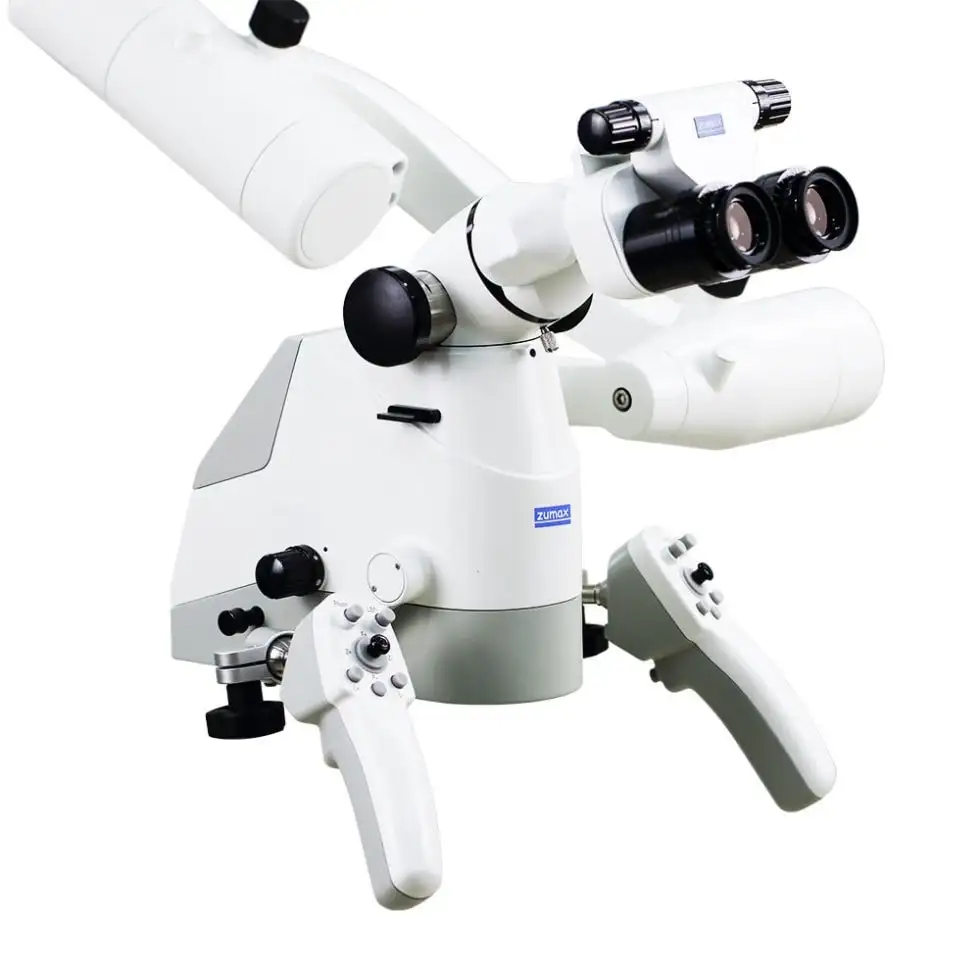
Magnification and focused light reveal details inside the tooth that the naked eye cannot see.
Why a microscope
Root canals are tiny, and some are hidden, calcified or curved. Under the microscope I see the canal entrances clearly, follow them more precisely, and check that the chamber is clean before filling.
Where it makes a difference
- Looking for an extra canal, such as the second mesiobuccal canal (MB2) in maxillary molars
- Retreating a previously treated tooth
- Revealing fine cracks
- Root-end microsurgery
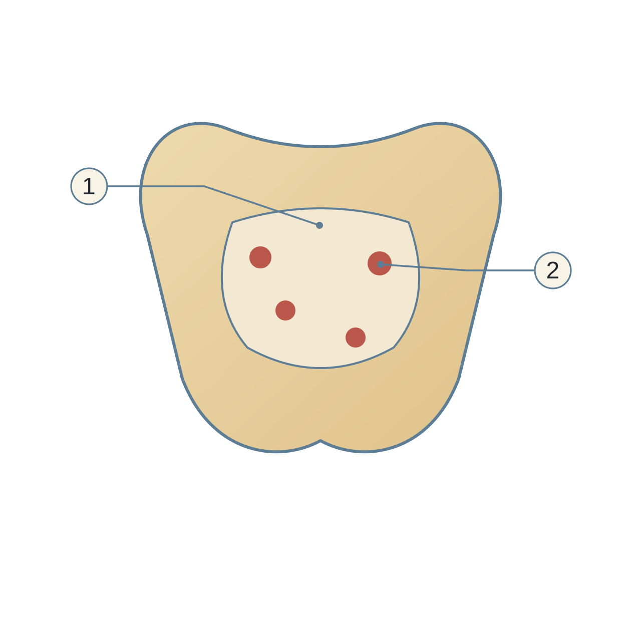
- Pulp chamber floor
A schematic view from above.
- Canal orifices
Their number and positions vary between teeth.
The microscope I use
I use a dental operating microscope, one of the latest microscope systems available.
A microscope supports clinical skill; it does not replace it, and it does not guarantee success.
Related reading


Core treatments

Medical notice: the information here is for general health education and is not a substitute for an examination and assessment by a dentist. If you notice any symptoms or changes in your teeth or gums, even if they seem minor, do not ignore them; consult a dentist to assess them and decide whether they need treatment.