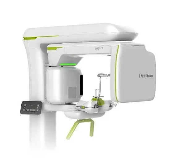
Cone-beam computed tomography (CBCT) provides three-dimensional images of the teeth and surrounding bone. It can be useful when the information from the examination and two-dimensional X-rays is not enough to make a treatment decision.
What can it show?
It can help assess the anatomy of the roots, periapical lesions, the location of some cases of root resorption and fractures, and planning endodontic microsurgery.
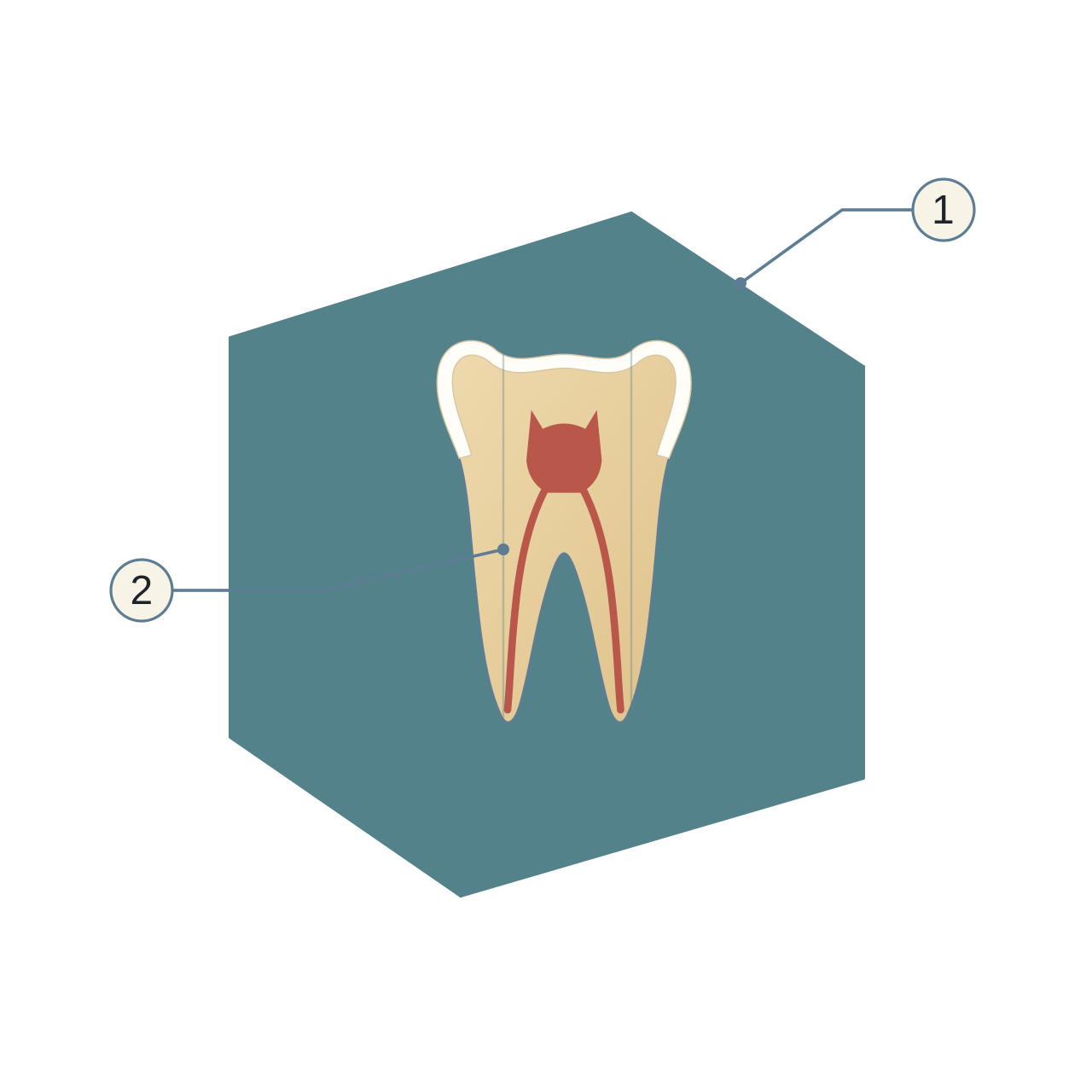
- Imaging volume
A simplified volume.
- Multiple sections
Help assess structures from different directions.
When do I use it?
It may be requested in specific cases such as complex retreatment, suspected fractures or root resorption, or when three-dimensional information is necessary to guide the treatment plan.
Does it replace ordinary X-rays?
No. Assessment starts with a clinical examination and the suitable imaging with the least radiation exposure, and CBCT is used when it is likely to add influential information that simpler methods do not provide.
What about the radiation dose?
The dose of CBCT may be higher than intraoral dental images, so suitable settings and the smallest field of view that meets the diagnostic need are chosen, with special care for children and adolescents.
The device I use
I use a CBCT system.
The takeaway
What makes CBCT useful is the additional information it can provide, which may change the diagnosis or the treatment decision, beyond the three-dimensional image itself.
I do not use it for every patient. It is requested when there is a clinical reason, with a small field covering only the area of interest to reduce the radiation dose.
Related reading


Core treatments


Medical notice: the information here is for general health education and is not a substitute for an examination and assessment by a dentist. If you notice any symptoms or changes in your teeth or gums, even if they seem minor, do not ignore them; consult a dentist to assess them and decide whether they need treatment.