A condition in which the body begins to dissolve part of a tooth root. It is usually painless and found on X-rays.

Internal and external
In internal resorption the walls of the canal are dissolved from within, often after chronic inflammation or injury. In external resorption it starts at the root surface after trauma, gum inflammation, orthodontic movement or pressure from an unerupted tooth.
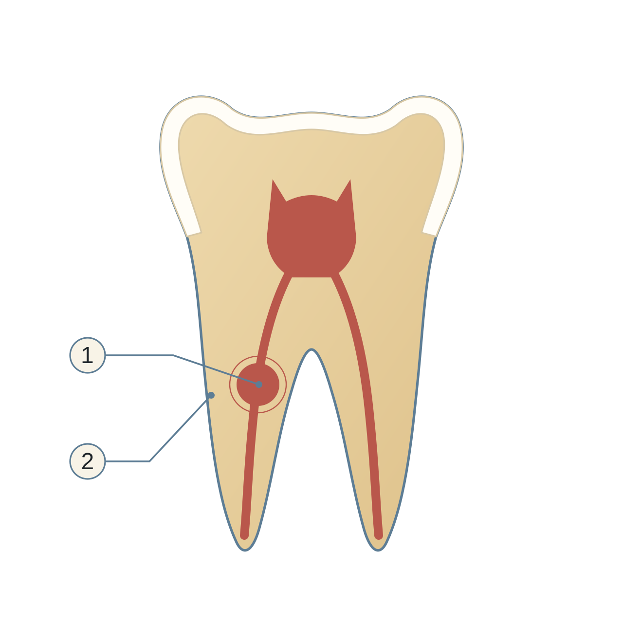
- Internal root resorption
Enlargement originating at the pulp-space wall.
- External root surface
No perforation is shown in this example.
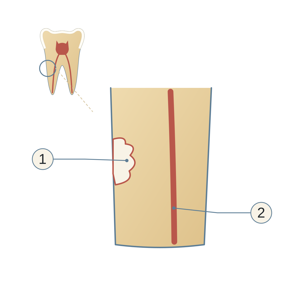
- External root resorption
An example of a defect starting at the root surface.
- Root canal
Not the centre of the illustrated defect.
Why precise diagnosis matters
The type and location decide whether the tooth can be saved and how. A standard two-dimensional X-ray may not show this clearly, so limited-field-of-view cone-beam computed tomography (CBCT) lets me see the root in three dimensions.
Treatment
In some early cases progress can be stopped with root canal treatment or a local repair using biocompatible materials. In more advanced cases saving the tooth may not be possible. I discuss this realistically after diagnosis.
Related reading


Core treatments


Medical notice: the information here is for general health education and is not a substitute for an examination and assessment by a dentist. If you notice any symptoms or changes in your teeth or gums, even if they seem minor, do not ignore them; consult a dentist to assess them and decide whether they need treatment.